-
-
-
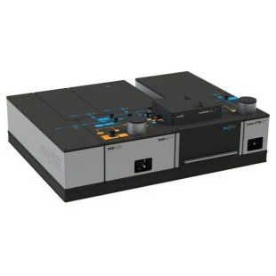
全自动外泌体荧光检测分析系统


- 品牌:NanoView
- 型号: 全自动外泌体荧光检测分析系统
- 产地:美国
- 供应商报价: 面议
-
QUANTUM量子科学仪器贸易(北京)有限公司
 更新时间:2024-04-18 22:55:56
 -
企业性质生产商
入驻年限第6年
营业执照已审核
- 同类产品成像系统(22件)
- 标签: 外泌体分析、外泌体荧光检测
联系方式:销售部010-85120280
联系我们时请说明在仪器网(www.yiqi.com)上看到的!
-
为您推荐


- 全自动外泌体荧光检测分析系统 核心参数
- 详细介绍
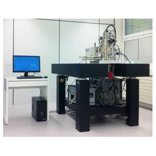
全自动外泌体荧光检测分析系统
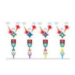
美国NanoView公司所开发的全自动外泌体荧光检测分析系统是一款无需纯化的、全自动的可对单个外泌体进行表征分析的全新设备。该设备能够提供全方位的外泌体表征信息,包括外泌体粒径大小、计数、分布、携带蛋白表达、生物标志物(CD9,CD81,CD63等)共定位等。操作简单,结果可靠。一经推出,便引起了外泌体领域科研工作者的广泛关注,短短两年时间在全·球已有50多个实验室采用该技术,发表重要文献近百篇。
全自动外泌体荧光检测分析系统的基本原理是一种基于特异性免疫捕获技术,允许研究者直接分析特定群体的外泌体或外囊泡。通过配套的试剂盒,客户一次性能够分析多达9个不同的样本,大大节省了时间和经济成本。全自动外泌体荧光检测分析系统兼容各种生物样本,除了纯化的外泌体之外,对于血液、尿液、恶性肿瘤、腹水中的外泌体也可直接检测分析,大大拓展了研究范围。
应用方向及主要特征

生物标记物共定位
可量化4种标记物的表达情况

计数分析
直接从样品中计算抗原阳性外泌体的数量,无需提纯

粒径分析
高精度统计外泌体的颗粒大小及分布

检测外泌体内容物
使用ExoView Cargo试剂盒可探测外泌体内部核酸的装载情况,分析装载率

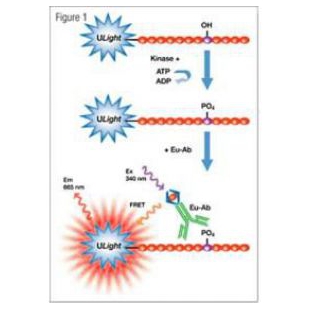
荧光检测
具备3个荧光通道,能够探测单个蛋白的结合

线性工作流程
9个样品的全自动分析

无须纯化
无需担心纯化带来的误差,更精确的测量样品间的表征和表达信息的差异

多重样本分析
可使用6种表面标记物来筛选外泌体
ExoView™ 参数信息:
- 颗粒大小分辨范围:大于50 nm(可分析大于40 nm的病毒颗粒)
- 荧光粒径分辨范围:大于20 nm
- 所需样本体积:25 μL
- 激发波长:410 nm,488 nm,555 nm,640 nm
- 可一次检测16个样本,每个样本可同时检测6个不同亚型及3种生物标记的荧光定位
- 单个样品检测时间:8分钟
- 捕获抗体:一个芯片多允许6种捕获抗体(+阴性对照)
- 荧光通道:3个荧光通道
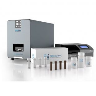
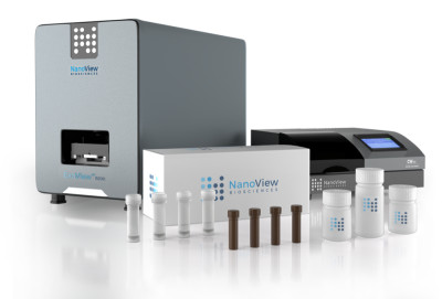
ExoView™ Kit 配置清单:
ExoView™
Tetraspanin Kits
CD9 - 捕获抗体
CD63 - 捕获抗体
CD81 - 捕获抗体
Mouse IgG 阴性对照
CD9、CD63、CD81三色共标记荧光抗体。
ExoView™
Tetraspanin Plasma Kits
CD9 - 捕获抗体
CD63 - 捕获抗体
CD81 - 捕获抗体
CD41a - 捕获抗体
多达三种定制化捕获抗体
同种阴性对照
CD9、CD63、CD81三色共标记荧光抗体。
ExoView™
Tetraspanin Custom Kits
CD9 - 捕获抗体
CD63 - 捕获抗体
CD81 - 捕获抗体
多达三种定制化捕获抗体
同种阴性对照
CD9、CD63、CD81三色共标记荧光抗体。
ExoView™
Cargo Kits
兼容所有ExoView Kit
标准 CD9, CD63和CD81 捕获抗体
Mouse IgG 阴性对照
ExoView Cargo 试剂
CD9、CD63、CD81三色共标记荧光抗体。
Sytenin荧光抗体
粒径分析
Exoview能够对>50 nm的外泌体进行全方面的表征,无论是粒径尺寸、粒径分布还是外泌体的亚型均可在一次测试中得到。并且所用来测试的样本无需进行纯化,避免因纯化带来的样本偏差。
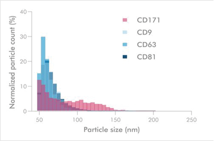
分析不同大小和不同尺度的外泌体亚群,在CD171过表达系统中,100 nm以上的外泌体仅表达了CD171。
量化外泌体亚群
通过抗体捕获的模式,Exoview能够量化含有不同标记物的外泌体亚群,并对不同种群的外泌体进行特异性计数和分析。整个过程无需纯化,并且线性范围可跨越3个数量级。

TSPAN8阳性细胞外囊泡的稀释曲线。使用1:3比例的梯度稀释。用R2 = 0.9986计算与TSPAN8荧光数据的线性拟合。
检测外囊泡中的装载物
MISEV建议在表征外泌体和外囊泡时,应当同时测量表面和载体蛋白。使用ExoView芯片可穿透外泌体,探测膜内蛋白和载体。并且单次可定量3种表面、管腔蛋白。
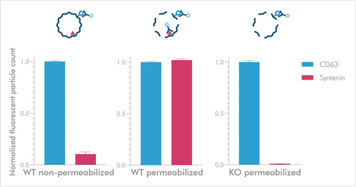
检测细胞外泌体内的Syntenin。
WT细胞在未穿膜的条件下几乎观测不到Syntenin的信号。进行穿膜处理后可以观测到Syntenin信号,而KO之后信号消失
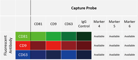
生物标志物共定位
ExoView能够在单个外泌体样本上检测多达4种标记物,并同时提供外泌体的其它表征数据诸如计数、颗粒尺寸统计等。
无需纯化
ExoView只测量含有靶标抗原的特定细胞外泌体群体。仅需35ul的稀释样品,即可直接获得外泌体的表型、l粒径和计数信息。并且无需担心污染物对测试的影响。
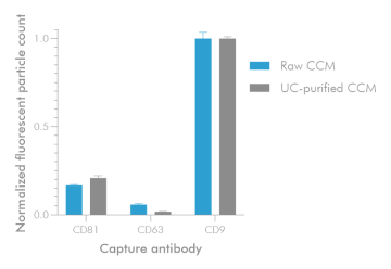
无论纯化与否,Exoview均可进行分析
相关视频
发表文章
2020年已发表文献
1. Cytokine profiling in serum-derived exosomes isolated by different methods, Jung HH, Kim JY, Lim JE, Im YH, Nature Scientific Reports 2020
2. Study of immune-tolerized cell lines and extracellular vesicles inductive environment promoting continuous expression and secretion of HLA-G from semiallograft immune tolerance during pregnancy, Cho K, Kook H, Kang S Lee J, JEV 2020
3. Annexin A1–dependent tethering promotes extracellular vesicle aggregation revealed with single–extracellular vesicle analysis, Rogers MA, Buffolo F, Schlotter F, Atkins SK, Lee LH, Halu A, Blaser MC, Tsolaki E Higashi H, Luther K, Daaboul G, Bouten CVC, Body SC, Singh SA, Bertazzo S, Libby P, Aikawa M, Aikawa E, Cell Biology 2020
4. Targeting tumor-derived exosomes using a lectin affinity hemofiltration device, Marleau AM, Jacobs MT, Gruber N, Rodell TC, Ferrone S, Whiteside TL, Cancer Research 2020
5. Extracellular Vesicle and Particle Biomarkers DefineMultiple Human Cancers. Hoshino A, Kim HS, Bojmar L, Gyan KE, Cioffi M, Hernandez J, Zambirinis CP, Rodriques G, Molina H, Heissel S, Mark MT, Steiner L, Benito Martin A, Lucotti S, Di Giannatale A, Offer K, Nakajima M, Williams C, Lyden D., Cell 2020
6. Reporter mice for isolating and auditing cell type‐specific extracellular vesicles in vivo, McCann JV, Bischoff SR, Zhang Y, Cowley DO, Sanchez-Gonzalez V, Daaboul GG, Dudley AC, Genesis 2020
7. High yield and scalable EV production from suspension cells triggered by turbulence in a bioreactor, Grainger A, Wilhelm C, Gazeuah F, Silva A, Cytotherapy 2020
8. Extracellular Vesicles: A New Frontier for Research in Acute Respiratory Distress Syndrome, Mahida RY, Matsumoto S, Matthay MA. American Journal of Respiratory Cell and Molecular Biology 2020
9. Small extracellular vesicles modulated by αVβ3 integrin induce neuroendocrine differentiation in recipient cancer cells, Quaglia F, Krishn SR, Daaboul GG, Sarker S, Pippa R, Domingo-Domenech J, Kumar G, Fortina P, McCue P, Kelly WK, Beltran H, Liu Q, Languino LR., Journal of Extracellular Vesicles, Volume 9, Issue 1, 2020
10. Characterisation of extracellular vesicles surface markers and co-expression studies with single particle interferometric imaging platform, Kusuma G, Lim R., Cytotherapy, Volume 22, Issue 5, 2020
11. Engineering mesenchymal stem cell paracrine activity with 3D culture, Kusuma G, LiA, Zhu D, McDonald H, Chmabers D, Frith J, Lim R.,Cytotherapy, Volume 22, Issue 5, 2020
12. Release of extracellular vesicle miR-494-3p by ARPE-19 cells with impaired mitochondria,Ahn JY, Datta S, Cano M, Mallick E, Rai U, Powell B, Tian J, Witwer KW, Handa JT, Paulaitis ME.,Biochim Biophys Acta Gen Subj. 2020 Mar 30:129598. (doi: 10.1016/j.bbagen.2020.129598)
13. Advantageous Antibody Microarray Fabrication Through DNA-Directed Immobilization: A Step Toward Use of Extracellular Vesicles in Diagnostics, Brambila D, Sola L, Chiari M., Talanta 2020
14. Unannotated small RNA clusters in circulating extracellular vesicles detect early stage liver cancer, Villanueva et al, bioRxiv preprint 2020 (doi: https://doi.org/10.1101/2020.04.29.066183)
15. Membrane-Binding Peptides for Extracellular Vesicles On-Chip Analysis, Gori A, Romanato A, Bergamaschi G, Strada A, Gagni P, Frigerio R, Brambilla D, Vago R, Galbiati S, PIcciolini S, Bedoni M, Daaboul G, Chiari M, Cretich M., Journal of Extracellular Vesicles, Volume 9, Issue 1, 2020
16. Subpopulations of extracellular vesicles from human metastatic melanoma tissue identified by quantitative proteomics after optimized isolation, Crescitelli R, L?sser C, Jang SC, Cvjetkovic A, Malmh?ll C, Karimi N, H??g JL, Johansson I, Fuchs J, Thorsell A, Gho YS, Bagge RO, L?tvall J, Journal of Extracellular Vesicles , 2020
17. Extracellular Vesicles Derived from Induced Pluripotent Stem Cells Promote Renoprotection in Acute Kidney Injury Model, Collino F, Lopes JA, Tapparo M, Tortelote GG, Kasai-Brunswick TH, Lopes GMC, Almeida DB, Skovronova R, Wendt CHC, de Miranda K, Bussolati B, Vieyra A, Soares Lindoso R., Cells, 2020
18. Extracellular Vesicles: A New Frontier for Research in Acute Respiratory Distress Syndrome,Mahida RY, Matsumoto S, Matthay MA. American Journal of Respiratory Cell and Molecular Biology, 2020
19. Exosomes A clinical COmpendium - Methods for exosome isolation and characterization,Zhou M, Weber SR, Zhao Y, Chan H, Sundstrom JM. Exosomes A Clinical Compendium 2020
20. Phenotypic analysis of extracellular vesicles: a review on the applications of fluorescence,Panagopoulou MS, Wark AW, Birch DJS, Gregory CD. Journal of Extracellular Vesicles, 2020
21. Immune Cell-Derived Exosomes in the Cancer-Immunity Cycle Yan W, Jiang S, Trends in Cancer 2020
- 产品优势
- 全自动外泌体荧光检测分析系统的基本原理是一种基于特异性免疫捕获技术,允许研究者直接分析特定群体的外泌体或外囊泡。通过配套的试剂盒,客户一次性能够分析多达9个不同的样本,大大节省了时间和经济成本。全自动外泌体荧光检测分析系统兼容各种生物样本,除了纯化的外泌体之外,对于血液、尿液、恶性肿瘤、腹水中的外泌体也可直接检测分析,大大拓展了研究范围。
- 产品文章
| 仪器分类: | 倒置 | 结构分类: | 透射式 |