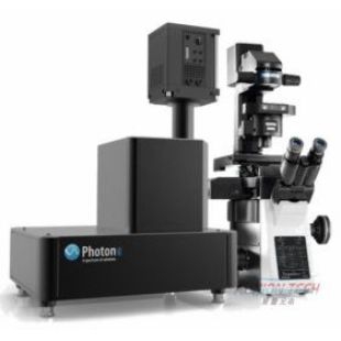
IMA荧光(EL/PL)显微高光谱成像仪
高速读取荧光高光谱
均化激光不会损伤细胞等样品
非逐点扫描
高速PL/EL Mapping
基于独特的体布拉格光栅滤波片技术(BTF)和光致发光成像技术,Photon etc公司zui新推出的IMA激光荧光显微高光谱成像系统,采取革新的二维成像的方式,激光经过扩束后再经过匀化,将高斯分布的点激光扩展成平面均匀分布面激光,面激光均匀照射在样品上,可以直接获得整个样品的荧光高光谱信息。从而获得分子结构方面的信息。有别于传统的激光荧光显微高光谱系统以逐点扫描的方式,而是一次性的获取整个样品的光谱信息,故而只需要更短的成像时间以及具有更高的空间分辨率。
共焦荧光成像系统,共聚焦荧光成像系统,共焦荧光光谱成像系统,共焦荧光成像光谱仪系统,成像光谱仪,激光荧光成像系统, 荧光显微成像系统
系统参数:
VIS | IR | |
光谱范围 | 400-1000nm | 900-1700nm |
光谱分辨率 | <2.5nm(最小可到0.2nm) | <4nm(最小可到0.4nm) |
图像分辨率 | 亚微米 | 亚微米 |
成像速度 | 20x20μm in 1s @100X | 20x20μm in 1s @100X |
激发光源 | 447 nm, 532, 635 nm(可选其他波长) | 808,980nm(可选其他波长) |
CCD | 科学级CCD,背照式CCD,EMCCD等 | InGaAs相机 |
显微镜 | 倒置或正置 | 倒置或正置 |
物镜 | 20X,60X,100X | 20X,60X,100X |
应用领域:
NANOPARTICLES IN CANCER CELLS
Dark field illumination is commonly used for the analysis of biological samples containing nanomaterials that significantly scatter light. When combined to hyperspectral imaging, it becomes an exceptional tool to also detect the composition and the location of nanomaterials embedded in cells. IMATM, Photon etc.’s hyperspectral imager, can be equipped with a highly efficient dark field condenser and generate high contrast images of biological samples.
The high throughput of Photon etc.’s hyperspectral filter allows the rapid acquisition of spectrally resolved high resolution images. Since the camera captures the whole area in the field of view, it is possible to collect spectral and spatial information in real time, with the possibility of recording spectrally resolved videos to follow the dynamics of cells and luminescent nanoscale components. PHySpecTM, Photon etc software, enables principal component analysis (PCA) in order to identify the smallest variations of single and aggregated nanoparticles.
With the purpose of showing the capabilities of IMATM to analyse nanomaterials in biological systems, a sample of MDA-MB-23 human breast cancer cells has been tagged with 60 nm gold nanoparticles (GNPs) and exposed to a dark field illumination on the entire field of view (Figure 1). With a 60×objective, an area of 150×112 μm was imaged, with a step of 2 nm and an exposition time of 2 s per wavelength. The complete analysis took only a few minutes, for more than one million spectra, each of them covering the whole visible spectrum.
Cells typically have a flat scattering spectrum, whereas GNPs show a sharp peak around 550 nm. Figure 2 illustrates the 550 nm image extracted from the dark field hyperspectral cube of the breast cancer. The GNPs are marked with a green colouring after PCA software processing. The magnification of a breast cancer cell (Figure 3a) and the spectra of the regions containing GNPs (some examples in Figure 3b) confirmed the presence of single 60 nm NPs (peak at 550 nm) and their aggregates (peaks red-shifted). The hyperspectral camera did not detect any GNPs in the areas between the cells.
CHARACTERISATION OF SOLAR CELLS USING HYPERSPECTRAL IMAGER
A new characterization method based on hyperspectral imaging recording spectrally resolved images allows the cartography of electroluminescence (EL) and photoluminescence (PL). From the data acquired, spatial variations of cell properties such as open circuit voltage and transport mechanisms were identified and characterized. Furthermore, the system was compared to a classical confocal microscope, showing significant gains in acquisition time.
Spectrally resolved images provide considerable advantages such as, absolute calibration of intensity, micrometer scale resolution, and excitation and detection on a surface (no information loss from lateral diffusion and roughness). In luminescence imaging, absolute calibration is a main concern and is here done in two steps: first, an absolute calibration at a determined point (spatially and spectrally) with a laser, and then a relative calibration on the whole space and the whole spectrum, with a calibrated lamp coupled to an integrating sphere.The images rendered by IMATM are spectrally resolved luminescence images from multicrystalline CIS solar cell, offering means of studying its spatial inhomogeneities. On high efficiency GaAs solar cells, we got absolute measurements of EL and successfully investigated reciprocity relations. Our next step is to record quantitative maps of CIGS physical properties from PL and EL images, such as VOC , transport parameters and more.
A confocal microscope coupled to a spectrometer provides similar data. The 532nm laser is focused onto the cell front contact, and the cartography of PL spectra is obtained by scanning the sample. The acquisition time with the imager is much faster. 150*150μm2 at 107 W/m2 would take hundreds of hours in confocal, but only 8min with IMA. Moreover, surface excitation and detection allow to get rid of diffusion and roughness troubles for quantitative analysis.
MULTIPLEXING OF 17 SWCNTs
NIR hyperspectral microscopy covers the detection range of 900-1700 nm and is ideal for the spatial and spectral identification and measurement of fluorophores that emit in the second biological window. For example, single wall nanotubes (SWNTs) emission bands are narrow (~ 20 nm) and each band corresponds to unique (n, m) species (chiralities). With IR hyperspectral microscopy, it is possible to separate these species, with single SWNT spatial resolution on surfaces, in live cells, and in vivo.
Images obtained by IR hyperspectral microscopy can be used to study fluorescence and spectral heterogeneity from single SWNTs in complex environments, including live cells and tissues.
Locate and identify single SWNTs chiralities
Identify SWNTs by their IR spectra
Separate single SWNT (emission band ~ 20 nm)
Simultaneous imaging of all emitters
Multiplexing with one laser source
Access to second biological window
Attenuated tissue absorbance
Higher depth of penetration
Less scattering
Limited autofluorescence
Monitor spectral information
Changes in intensity of single emitters
Shifts in wavelength
Spectral bandwidth variations
In vivo applications
In vivo imaging of multiplexed emitters
In vivo long term sensing
- 产品分类
- 品牌分类
- 冷原子/量子光学产品
- 位移台/偏转镜/隔振台
- 光纤/光纤器件
- 晶体、光栅
- 光学器件
- 光谱仪器
- 信号处理与分析设备
- 空间光调制产品
- 调制器/偏转器/滤波器
- 激光量测设备
- 相机系列
- 脉冲激光器(ns/ps/fs)
- 连续激光器(CW)
- THz产品
- X射线产品
- 材料分析设备
- 光学检测设备
- 其他专业应用
- 科研/工业成像系统
- 激光共聚焦显微拉曼-荧光光谱成像系统
- 激光扫描光电流显微成像系统
- 显微成像系统及组件
- 光学隔振平台
- 可调谐激光器
- 电子学测试仪器
- 可调谐光源 | 宽带光源
- (法国)法国Oxxius
- (美国)美国REO
- (德国)德国Asphericon
- (加拿大)加拿大Photon etc
- (俄罗斯)俄罗斯TekhnoScan
- (德国)德国GOM
- (法国)法国Cristal Laser SA
- (法国)法国BKtel Photonics
- (德国)德国quTools
- (法国)法国PHOTONICS BRETAGNE
- (法国)法国Manlight
- (美国)美国Double Helix
- (匈牙利)匈牙利CE Optics
- (美国)美国Calmar
- (德国)德国西克
- (美国)美国Optromix QSY
- (俄罗斯)俄罗斯阿维斯塔
- (德国)奥地利Xarion
- (瑞士)瑞士苏黎世仪器
- (瑞士)瑞士NKT Photonics
- (美国)美国Semiconsoft
- (日本)日本NEOARK
- (法国)法国Leukos
- (法国)法国PIEZOCONCEPT
- (法国)法国GLOphotonics
- (美国)美国Swamp Optics
- (日本)日本Artray
- (美国)美国MinusK
- (法国)法国Qiova
- (西班牙)西班牙NIT
- (加拿大)加拿大INO
- (德国)德国greateyes
- (法国)法国iXblue
- (日本)日本Meiritz
- (其它)爱沙尼亚beecher
- (美国)美国Teledyne Hastings
- (德国)德国Greateye
- (深圳)深圳大疆
- (德国)德国XImea
- (美国)美国Chroma
- (德国)德国Cycle
- (德国)德国Sciospec
- (瑞士)瑞士Arcoptix
- (美国)美国菲力尔
- (美国)美国ALIO
- (挪威)挪威Zivid
- (比利时)比利时IMEC
- (美国)美国必达泰克
- (澳大利亚)澳大利亚MOGLabs
- (瑞士)瑞士彩虹光电
- (瑞士)瑞士SENIS AG
- (芬兰)芬兰Spectral Engines
- (德国)德国Knick
- (立陶宛)立陶宛Quantum Light Ins
- (德国)德国PROTECT
- (英国)英国ForthDD
- (意大利)意大利Elements
- (立陶宛)立陶宛OptoGama
- (法国)法国AUREA
- (德国)德国Avenir Photonics
- (荷兰)荷兰Admesy
- (芬兰)芬兰SPECIM
- (法国)SPARK LASERS
- (美国)美国Quantum-Si
- (德国)德国CeramOptec
- (其它)白俄罗斯SOLAR LS
- (德国)Qioptiq
- (意大利)意大利Optogenix
- (德国)德国TEM Messtechnik
- (美国)美国莱特浩斯
- (徐汇区)昊量/auniontech
- (意大利)意大利Active Technologies
- (法国)法国IXFiber
- (昌平区)北京普源
- (美国)Pranalytica
- (日本)日本Kimmon
- (美国)美国New Scale Technologies
- (美国)美国TMC
- (意大利)意大利Bright Solutions
- (英国)英国Covesion
- (美国)美国Herzan
- (美国)美国OptiGrate
- (法国)法国Silios
- (澳大利亚)澳大利亚Liquid
- (美国)Vixar
- (美国)美国CTI
- (德国)德国alphalas
- (德国)德国TEM
- (法国)法国Alpao
- (比利时)比利时Xenics
- (美国)美国Hinds
- (日本)日本吉奥马
- (德国)德国PCO
- (美国)美国Meadowlark Optics
- (英国)英国ForthDD
- (德国)德国Yuanda Tech
- (德国)德国Cinogy
- (德国)德国VIALUX
- (意大利)意大利El.En.
- (英国)英国Gooch&Housego
- (法国)法国Phasics
- (无锡)无锡布里渊
- (德国)德国INSION
- (法国)法国Resolution Spectra System
- (俄罗斯)俄罗斯OSTEC
- (韩国)韩国Nanobase
- (美国)美国BNS
- (美国)美国Photo Research
- (俄罗斯)俄罗斯Scontel
- (法国)法国Photline Technologies
- (美国)美国Stable Laser Systems
- (美国)美国ConOptics
- (法国)法国ISP
- (德国)德国Gigahertz-Optik
-
仪企号
 上海昊量光电设备有限公司
-
友情链接