-
 银牌会员 第 9 年
QUANTUM量子科学仪器贸易(北京)有限公司
认证:工商信息已核实
- 产品分类
- 品牌分类
- ( 美国)美国Lake Shore
- ( 英国)英国iotaSciences
- ( 德国)德国REACNOSTICS
- ( 美国)MONSTR Sense
- ( 瑞典)Viventis Microscopy
- ( 西班牙)NEXT-TIP
- ( 加拿大)ICSPI
- ( 法国)Spin-ION
- ( 法国)塔莱恩特(Telight)
- ( 美国)zeroK Nanotech
- ( 德国)Kiutra
- ( 美国)NanoView
- ( 青岛)致真精密仪器
- ( 西班牙)Planelight
- ( 英国)Moorfield
- ( 瑞士)Qzabre
- ( 西班牙)Das Nano
- ( 波兰)Novilet
- ( 日本)GES
- ( 美国)美国PHOTOTHERAML
- ( 美国)美国Applied NanoFluorescence
- ( 日本)日本Tohuko
- ( 法国)法国Alyxan
- ( 美国)美国Anasazi
- ( 日本)日本Churitsu
- ( 日本)日本RIBM
- ( 德国)德国LLS ROWIAK
- ( 西班牙)西班牙Nanoscale Biomagnetic
- ( 英国)英国ONI
- ( 美国)美国Mizar Imaging
- ( 荷兰)荷兰Lumicks
- ( 瑞士)瑞士Cytosurge
- ( 加拿大)加拿大Johnsen Ultrava
- ( 美国)美国Depths of the eart
- ( 美国)美国Thermal Technology
- ( 韩国)韩国NTi
- ( 美国)美国BlueWave
- ( 德国)德国SciDre
- ( 瑞士)瑞士Swisslitho AG
- ( 美国)美国ARRADIANCE
- ( 奥地利)奥地利SCL-Sensor.Tech
- ( 日本)Nanophoton
- ( 奥地利)奥地利GETec
- ( 芬兰)芬兰SPECIM
- ( 美国)美国RHK Technology
- ( 美国)美国Delong Instruments
- ( 日本)PULSTEC
- ( 瑞士)瑞士IRsweep
- ( 德国)德国neaspec
- ( 美国)美国easyXAFS
- ( 德国)德国PANCO
- ( 日本)日本Advance Riko
- ( 德国) 德国LOT-Orial Group
- ( 法国)法国Hprobe
- ( 比利时)比利时Metis
- ( 瑞典)瑞典NanOsc
- ( 英国)英国Durham Magneto Optic
- ( 美国)美国MicroSense
- ( 美国)美国HPD
- ( 英国)英国ICEoxford
- ( 瑞典)Excillum
- ( 德国)德国THEVA
- ( 德国)德国Attocube Systems
- ( 美国)美国Montana Instruments
- ( 美国)美国Quantum Design
- ( 瑞士)attolight
- ( 法国)法国abbelight
- ( 日本)Omegatron
- ( 浦东新区)Nanoink
- ( 浦东新区)Cyberstar
- ( 浦东新区)Montana
- ( 德国)NanoTemper
- ( 德国)Nanoanalytics
- ( 浦东新区)ARRADIANCE
- ( 德国)iplas
- ( 浦东新区)MicroSense
- ( 浦东新区)HPD
- ( 浦东新区)Cyberstar
- ( 浦东新区)Thermal Technology
- ( 浦东新区)Alyxan
-
仪企号
 Quantum Design中国子公司


-
-
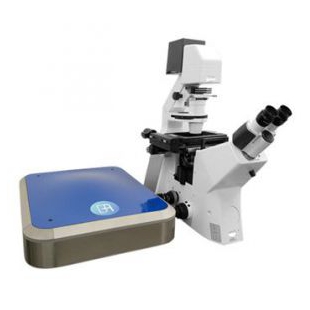
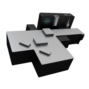
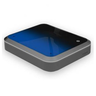
- 品牌:塔莱恩特(Telight)
- 型号:LiveCodim
- 产地:欧洲 法国
- 供应商报价:面议
| 品牌: | 塔莱恩特(Telight) | 型号: | LiveCodim |
模块化超分辨共聚焦显微系统-LiveCodim
传统荧光显微镜受到光学衍射极限的影响,高的分辨率为200 nm,因此很难观察细胞中的超微结构。LiveCodim是一款模块化超分辨共聚焦显微系统,能够适配绝大多数的倒置荧光显微镜,将现有的倒置显微镜升级成为具备宽场、共聚焦、超分辨三大模式的成像系统。LiveCodim通过独特的锥形衍射显微镜—— 一种强大的波束成形器,能够直接提供分辨率高达120 nm的实时活细胞超分辨共聚焦成像,同时无需对样品进行任何额外操作,结合其低光毒性,以及方便快捷的操作系统等优势,非常适合拍摄荧光成像。
产品优势
· 超高性价比:模块化超分辨,节省成本,兼容绝大多数倒置显微镜
· xy轴超高分辨率:<120 nm
· z轴深度成像:具备z-stack成像能力,高成像深度50 μm
· 活细胞成像:低光毒性和光漂白性,适合活细胞成像
· 制样简单:样品无需特殊处理,无需特殊染料
· 全自动软件:全自动调节各种参数,简单易上手
主要参数
· xy轴分辨率:< 120 nm
· z轴分辨率:< 500 nm
· z轴成像深度:50 μm
· 成像视野:共聚焦模式下80 μm * 80 μm,超分辨模式下: 50 μm * 50 μm
· 成像模式:宽场、共聚焦、LiveCodim超分辨
· 四色成像通道:405 nm, 488 nm, 561 nm, 640 nm (根据需求可增加)
测试数据
1. MDCK细胞中线粒体的动态变化

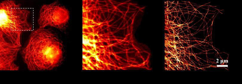
2. Hela胞的微管宽场,共聚焦,LiveCodim超分辨成像
3. 细胞分裂中期的COS-7细胞3D多色超分辨成像
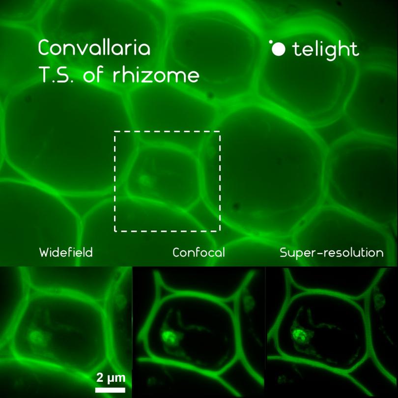
4. 植物细胞成像:观测铃兰草的根茎
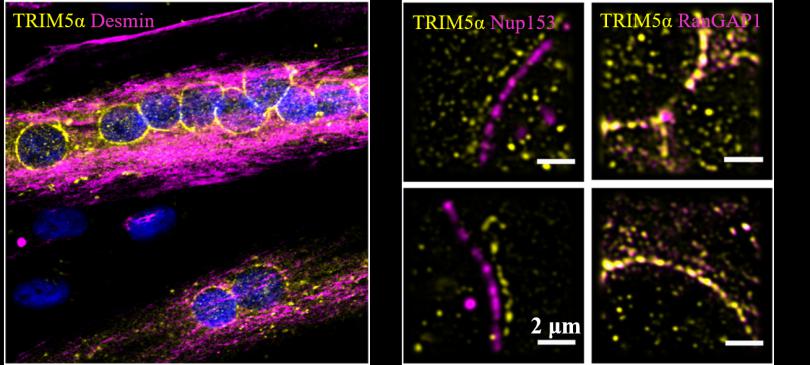
5. 天然免疫分子TRIM5α作用机制研究
天然免疫分子TRIM5α蛋白是人类基因中决定疾病的易感性和发病速度的重要因素,其抗病毒活性通常通过小泛素相关修饰物(SUMO)调节,但是具体的作用机制仍有待进一步研究。LiveCodim超分辨图像揭示了TRIM5α主要分布在肌小管的核膜上,同时与存在于核孔的细胞质丝上的RanGTPase激活蛋白RanGAP1有明显的共定位现象,和主要定位于核篮上的蛋白Nup153无明显共定位,说明TRIM5α主要定位于这类细胞的胞质面。
部分发表文章
[1] Fernandez, Juliette, et al. "Transportin-1 binds to the HIV-1 capsid via a nuclear localization signal and triggers uncoating." Nature microbiology 4.11 (2019): 1840-1850.
[2] Vargas, Jessica Y., et al. "The Wnt/Ca2+ pathway is involved in interneuronal communication mediated by tunneling nanotubes." The EMBO journal 38.23 (2019): e101230.
[3] Maarifi, Ghizlane, et al. "RanBP2 regulates the anti-retroviral activity of TRIM5α by SUMOylation at a predicted phosphorylated SUMOylation motif." Communications biology 1.1 (2018): 1-11.
[4] Garita-Hernandez, Marcela, et al. "Optogenetic light sensors in human retinal organoids." Frontiers in neuroscience 12 (2018): 789.
[5] Getz, Angela M., et al. "Tumor suppressor menin is required for subunit-specific nAChR α5 transcription and nAChR-dependent presynaptic facilitation in cultured mouse hippocampal neurons." Scientific reports 7.1 (2017): 1-16.
[6] Portilho, Débora M., Roger Persson, and Nathalie Arhel. "Role of non-motile microtubule-associated proteins in virus trafficking." Biomolecular concepts 7.5-6 (2016): 283-292.
[7] Pagliuso, Alessandro, et al. "A role for septin 2 in Drp1‐mediated mitochondrial fission." EMBO reports 17.6 (2016): 858-873.
[8] Fallet, Clement, and Gabriel Y. Sirat. "Achromatization of conical diffraction: application to the generation of a polychromatic optical vortex." Optics letters 41.4 (2016): 769-772.
[9] Fallet, Clement, et al. "Accurate axial localization by conical diffraction beam shaping generating a dark-helix PSF." Single Molecule Spectroscopy and Superresolution Imaging IX. Vol. 9714. International Society for Optics and Photonics, 2016.
[10] Fallet, Clement, Arvid Lindberg, and Gabriel Y. Sirat. "Generating 3D depletion distribution in an achromatic single-channel monolithic system." Single Molecule Spectroscopy and Superresolution Imaging IX. Vol. 9714. International Society for Optics and Photonics, 2016.
[11] Fallet, Clément, et al. "A new method to achieve tens of nm axial super-localization based on conical diffraction PSF shaping." Single Molecule Spectroscopy and Superresolution Imaging VIII. Vol. 9331. International Society for Optics and Photonics, 2015.
[12] Caron, Julien, et al. "Conical diffraction illumination opens the way for low phototoxicity super-resolution imaging." Cell adhesion & migration 8.5 (2014): 430-439.
[13] Fallet, Clément, et al. "Conical diffraction as a versatile building block to implement new imaging modalities for superresolution in fluorescence microscopy." Nanoimaging and Nanospectroscopy II. Vol. 9169. International Society for Optics and Photonics, 2014.
[14] Rosset, Sybille, Clement Fallet, and Gabriel Y. Sirat. "Focusing by a high numerical aperture lens of distributions generated by conical diffraction." Optics letters 39.23 (2014): 6569-6572.
用户单位

法国巴斯德研究所

蒙彼利埃大学