-
-
-
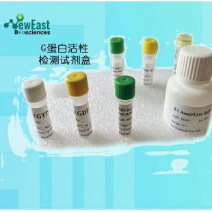
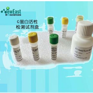
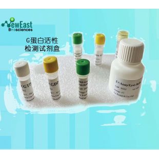
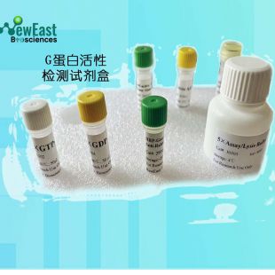
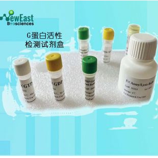
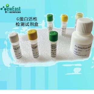
Arf6 活性检测试剂盒/Arf6 assay kit 现货推荐
- 品牌:NewEast Biosciences
- 型号/货号: 30Assays/82401
- 产地:湖北 武汉
- 供应商报价:面议
-
武汉费斯德生物科技有限公司
 更新时间:2023-04-16 23:55:39
 -
销售范围售全国
入驻年限第3年
营业执照已审核
- 同类产品活性抗体试剂盒(13件)

立即扫码咨询
联系方式:400-822-6768
联系我们时请说明在仪器网(www.yiqi.com)上看到的!
扫 码 分 享 -
为您推荐

产品特点
- 活性G蛋白检测试剂盒是基于特异性识别与 GTP 结合的,而不认识 GDP 结合的GTP酶的单克隆抗体。鉴于单克隆抗体对GTP酶的高亲和力,可以在短时间内进行活化测定。该试剂盒提供了具有高度灵敏性和可靠性。
详细介绍
Arf6 Pull-Down Activation Assay Kit
Cat. # 82401
Introduction
A. Background
ARF6 (ADP-ribosylation factor 6) is a member of the ARF super-family. ARF genes encode small GTPases that increase the ADP-ribosyltransferase activity of cholera toxin and are critical for vesicular trafficking as activators of phospholipase D. Arf6 regulates membrane trafficking and functions as a regulatory molecule of phagocytosis.Currently there is no direct assay to measure the activation of Arf6 GTPase.B. Assay Principle
The Arf6 Activation Assay Kit uses configuration-specific anti-Arf6-GTP Mouse monoclonal antibody to measure Arf6-GTP levels in cell extracts or in vitro GTPγS loading Arf6 activation assays. Anti-Arf6-GTP mouse monoclonal antibody is first incubated with cell lysates containing Arf6-GTP. Next, the GTP-bound Arf6 is pulled down by protein A/G agarose. Finally, the precipitated Arf6-GTP is detected through immunoblot analysis using Anti-Arf6 Rabbit Polyclonal Antibody.The anti-Arf6-GTP monoclonal antibody can also be used to monitor the activation of Arf6 in cells and in tissues by immunohistochemistry.C. Kit Components
1. Anti-Arf6-GTP Mouse Monoclonal Antibody (Cat. # 26918): One vial – 35 µL (1 mg/ml) in PBS, pH 7.4, containing 50% glycerol. This antibody specifically recognizes Arf6-GTP from all vertebrates.2. Protein A/G Agarose (Cat. # 30301): One vial – 600 µL of 50% slurry.3. 5X Assay/Lysis Buffer (Cat. # 30302): One bottle – 30 mL of 250 mM Tris-HCl, pH 8, 750 mM NaCl, 50 mM MgCl2, 5 mM EDTA, 5% Triton X-100.4. Anti-Arf6 Rabbit Polyclonal Antibody (Cat. # 21032): One vial – 50 µL (1 mg/mL) in PBS, pH 7.4, contained 50% glycerol.5. 100X GTPγS (Cat. # 30303): One vial – 50 µl at 10 mM, use 5 µL of GTPγS for GTP-labeling of 0.5 mL of cell lysate.6. 100X GDP (Cat. # 30304): One vial – 50 µl at 100 mM, use 5 µL of GDP for GDP-labeling of 0.5 mL of cell lysate.7. HRP-Goat Anti-Rabbit IgG (Cat. #29002): 50 µL (0.4 mg/mL) in PBS, pH 7.4, contained 50% glycerol.D. Materials Needed but Not Supplied
1. Stimulated and non-stimulated cell lysates2. Protease inhibitors3. 4 °C tube rocker or shaker4. 0.5 M EDTA at pH 8.05. 1.0 M MgCl26. 2X reducing SDS-PAGE sample buffer7. Electrophoresis and immunoblotting systems8. Immunoblotting wash buffer such as TBST (10 mM Tris-HCl, pH 7.4, 0.15 M NaCl, 0.05% Tween-20)9. Immunoblotting blocking buffer (TBST containing 5% Non-fat Dry Milk or 3% BSA)10. ECL Detection ReagentsE. Example Results
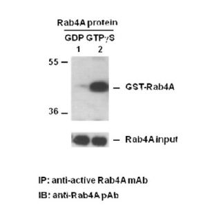
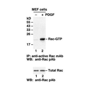
The following figure demonstrates example results seen with the Arf6 Activation Assay Kit. For reference only. Arf6 Activation Assay. Purified Arf6 proteins were immunoprecipitated after treated with GDP (lane 1) or GTPγS (lane 2). Immunoprecipitation was done with the anti-Arf6-GTP monoclonal antibody (Cat. No. 26918). Immunoblot was with an anti-Arf6 rabbit polyclonal antibody (Cat. No. 21032).
Assay Procedure
A. Reagent Preparation1X Assay/Lysis Buffer: Mix the 5X Stock (Cat. # 30301) briefly and dilute to 1X in deionized water. Just prior to usage, add protease inhibitors such as 1 mM PMSF, 10 µg/mL leupeptin, or 10 µg/mL aprotinin.B. Sample PreparationAdherent Cells
1. Culture cells (one 10-cm plate, ~107 cells) to approximately 80-90% confluence. Stimulate the cells with activator or inhibitor as desired.2. Aspirate the culture media and wash twice with ice-cold PBS.3. Completely remove the final PBS wash and add ice-cold 1X Assay/Lysis Buffer (See Reagent Preparation) to the cells (0.5-1 mL per 10 cm tissue culture plate).4. Place the culture plates on ice for 10-20 minutes.5. Detach the cells from the plates by scraping with a cell scraper.6. Transfer the lysates to appropriate size tubes and place on ice.7. If nuclear lysis occurs, the cell lysates may become viscous and difficult to pipette. If this occurs, lysates can be passed through a 27½-gauge syringe needle 3-4 times to shear the genomic DNA.8. Clear the lysates by centrifuging at 12,000 x g and 4°C for 10 minutes.9. Collect the supernatant and store the sample (~1-2 mg of total protein) on ice for immediate use, or snap freeze and store at -70°C for future use.Adherent Cells
1. Culture cells and stimulate with activator or inhibitor as desired.2. Perform a cell count and then pellet the cells through centrifugation.3. Aspirate the culture media and wash twice with ice-cold PBS.4. Completely remove the final PBS wash and add ice-cold 1X Assay/Lysis Buffer (See Reagent Preparation) to the cell pellet (0.5-1 mL per 107 cells).5. Lyse the cells by repeated pipetting.6. Transfer the lysates to appropriate size tubes and place them on ice.7. If nuclear lysis occurs, the cell lysates may become viscous and difficult to pipette. If this occurs, lysates can be passed through a 27½-gauge syringe needle 3-4 times to shear the genomic DNA.8. Clear the lysates by centrifuging at 12,000 x g and 4°C for 10 minutes.9. Collect the supernatant and store sample on ice for immediate use, or snap freeze and store at -70°C for future use.C. In vitro GTPγS/GDP Protein for Positive and Negative controls
Note: In vivo stimulation of cells will activate approximately 10% of the available Arf6, whereas in vitro GTPγS protein loading will activate nearly 90% of Arf6.1. Aliquot 0.5 mL of cell extract (or 1 µg of purified Arf6 protein) into two microcentrifuge tubes.2. To each tube, add 20 µL of 0.5 M EDTA (final concentration of 20 mM).3. Positive control: add 5 µL of 100 X GTPγS (Cat. # 30302) to the 1st tube4. Negative control: add 5 µL of 100 X GDP (Cat. # 30304) to the 2nd tube.5. Incubate both tubes at 30°C for 30 minutes with agitation.6. Stop loading by placing the tubes on ice and adding 32.5 µL of 1 M MgCl2 (final concentration of 60 mM).D. Affinity Precipitation of Activated G Protein
1. Aliquot 0.5-1 mL of cell lysates (about 1 mg of total cellular protein) to a microcentrifuge tube.2. Adjust the volume to 1 mL with 1X Assay/Lysis Buffer (See Reagent Preparation).3. Add 1 µL anti-Arf6-GTP antibody (Cat. # 26918).4. Prepare the protein A/G Agarose bead slurry (Cat. # 30301) by resuspending through vertexing or titrating.5. Quickly add 20 µL of resuspended bead slurry to above tube.6. Incubate the tube at 4°C for 1 hour with gentle agitation.7. Pellet the beads through centrifugation at 5,000 x g for 1 min.8. Aspirate and discard the supernatant (making sure not to disturb or remove the bead pellet.9. Wash the beads 3 times with 0.5 mL of 1X Assay/Lysis Buffer, centrifuging and aspirating each time.10. After the third wash, pellet the beads through centrifugation and carefully remove all the supernatant.11. Resuspend the bead pellet in 20 µL of 2X reducing SDS- PAGE sample buffer.12. Boil the sample for 5 minutes.13. Centrifuge it at 5,000 x g for 10 seconds.E. Western Blot Analysis
1. Load 15 µL/well of pull-down supernatant to a polyacrylamide gel (17%). It is recommended to include a pre-stained MW standard (as an indicator of a successful transfer in step 3 below).2. Perform SDS-PAGE following the manufacturer’s instructions.3. Transfer the gel proteins to a PVDF or nitrocellulose membrane following the manufacturer’s instructions.Note: Steps 4-11 are at room temperature with agitation4. Following electroblotting, immerse the PVDF membrane in 100% Methanol for 15 seconds, and then allow it to dry at room temperature for 5 minutes.Note: If Nitrocellulose is used instead of PVDF, step 4 Should be skipped.5. Block the membrane with 5% non-fat dry milk or 3% BSA in TBST for 1 hr at room temperature with constant agitation.6. Wash the blotted membrane three times with TBST, 5 minutes each time.7. Incubate the membrane with Anti-Arf6 Rabbit Polyclonal Antibody (Cat. # 21032), which is freshly diluted 1: 50~500 (depending on the amount of Arf6 proteins in your sample) in 5% non-fat dry milk or 3% BSA in TBST, for 1-2 hr at room temperature with constant agitation or at 4°C overnight.8. Wash the blotted membrane three times with TBST, 5 minutes each time.9. Incubate the membrane with a secondary antibody (Cat. # 29002), which is freshly diluted 1: 1000 in 5% non-fat dry milk or 3% BSA in TBST, for 1 hr at room temperature with constant agitation.10. Wash the blotted membrane three times with TBST, 5 minutes each time.11. Use the detection method of your choice such as ECL.
您可能感兴趣的产品
-
Arf6 活性检测试剂盒/Arf6 assay kit 现货推荐
-
 Rab3 活性检测试剂盒/NeweastBio现货更新推荐
-
 Rab35 活性检测试剂盒/NeweastBio产品推荐
-
 DNase Viability Assay Kit (Fluorescent Labeling) DNase活性检测试剂盒
-
 Arl1 活性检测试剂盒/2023现货试剂盒
-
 武汉试剂盒现货 Gα13活性检测试剂盒
-
 Rab11 活性检测试剂盒/NeweastBio现货采购
-
 Gαs 活性检测试剂盒/2023推荐商家更新
-
 蛋白Recombinant Human ARF6 protein (His tag)|Recombinant Human ARF6 protein (His tag)|Elabscience/伊莱瑞特|100μg
-
蛋白Recombinant Human ARF6 protein (His tag)|Recombinant Human ARF6 protein (His tag)|Elabscience/伊莱瑞特|20μg
-
 Catalase Assay Kit 过氧化氢酶检测试剂盒
-
ARF6 多克隆抗体|Elabscience/伊莱瑞特|20μL